This year, the Nobel Prize for Medicine is awarded to a branch of the discipline whose roots lie in a sophisticated spectroscopic technique usually the preserve of chemists, but which relies on the properties of the very superconducting magnets revealed in this year’s Physics Prize.
Indeed, the two scientists who share the Prize are physicist Peter Mansfield (70) of the University of Nottingham and chemist Paul Lauterbur (74), of the University of Illinois at Urbana-Champaign. The application of their work, however, is certainly of great benefit to medicine – magnetic resonance imaging.
Paul Lauterbur (Credit: Photo by Bill Wiegand)
Imaging the interior of the human body non-invasively was until just a few years ago reliant on X-rays, but soft tissues, such as kidneys, brains, and hearts were not apparent to such probing. Lauterbur and Mansfield’s work led to the development of a technique that has revolutionised diagnostics, treatment and follow-up.
Atomic nuclei held in a strong magnetic field rotate with a frequency that is dependent on the strength of the magnetic field. However, their total energy can be increased if they are also bathed in radio waves at the resonant frequency. When they relax back down to their previous energy level, they release the radio waves but at a different frequency. This seminal discovery was made in the 1950s and was recognised by the 1952 Nobel Prize for Physics.
Paul Lauterbur
Over the following years, this technique of magnetic resonance was developed into a powerful analytical tool for chemistry. By scanning a molecule with a sweep of radio waves it is possible to record the resonances of each atom in a molecule. Atoms in different positions emit radio waves at different shifts depending on the other atoms surrounding them. At the beginning of the 1970s, this year’s Nobel physicists made pioneering contributions to the development of this technique for scanning the atoms in solid objects in such a way that a two-dimensional slice through the object could be imaged.
Peter Mansfield
The human body is two thirds water, but different organs contain different amounts. By sweeping the hydrogen atoms in the water molecules with the radio waves of a magnetic resonance machine, it is possible to reveal these differences which are then correlated with organ structure and different tissues and converted into a 2D image of the organ. Computer control and interpretation allow parts of the body to be scanned and a series of slices recorded, which are then composed into a three-dimensional image. In many diseases, water content of an organ changes and this is reflected in the image. For instance, a solid tumour will have a much lower water content than surrounding healthy tissue in the brain, for instance, and will be clearly visible in the MR image.
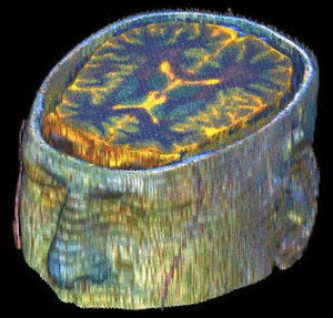
The head scan (Credit: J. Lambert/NASA)
MRI is especially suited to imaging the brain and the spinal cord where investigative surgery has become a last resort for many conditions from tumours to herniated discs. It also reveals the localised inflammation of multiple sclerosis and can be used in follow-up to treatment to visualise response to treatment with fine detail.
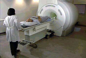
Scanner (Credit: GE Medical Systems Europe)
Magnetic resonance imaging, MRI, is now routine in countless hospitals, with some 60 million scans being carried out every year. The technique is still being voraciously developed by researchers hungry to improve on even the present powerful machines. Mansfield and his colleagues have, for instance, developed a spin-off technique known as Echo-Planar Imaging (EPI). Where MRI scans a line at a time, EPI takes a snapshot of the 2D slice in one go. This allows rapid changes in an organ to be visualised as well as avoiding the blur associated with a patient’s movements during the scan.
As ever, the Nobel Prize is not without controversy. Physician Raymond Damadian pioneered the application of NMR in medicine during the early 1970s working with rat tumours. His work predates that of Lauterbur and Mansfield. Indeed, Damadian built the first MRI scanner. However, this year’s winners used magnetic field gradients which led to the form of imaging hospitals now use routinely.
Further reading
Suggested searches
magnetic resonance imaging