Researchers have shown that networks of chitin filaments are an integral part of the silica shells of tiny marine creatures known as diatoms. The discovery could open the way to emulating these incredibly diverse and potentially very useful substances for materials science and engineering applications.
Some of the most bizarre and beautiful structural forms in nature exist in the world of the diatom, so named from the Greek meaning “cut through”. They represent a major group of eukaryotic algae that has existed since Jurassic times and are among the most common plant-type plankton, phytoplankton. They are mostly unicellular but form ribbon and fan-like colonies. Their most outstanding feature is that each cell is encapsulated by a unique shell of hydrated silicon dioxide, known as a frustule.
There is an almost infinite diversity of frustules but they commonly consist of two asymmetrical sides with a division down the middle, hence the name – diatom. Diatom frustules are not composed solely of hydrated silica but contain other substances, making them among the most fascinating natural hybrid materials having some quite unusual mechanical and optical properties.
However, scientists do not yet fully understand the biochemical mechanisms by which diatoms lay down silica and combine it with other substances in the biomineralization process of frustule construction.
Now, researchers at TU Dresden and the Max Planck Institute the Chemical Physics of Solids in Dresden, Germany, have identified another of the critical components of the diatom frustule. TU Dresden’s Eike Brunner and colleagues report details in the journal Angewandte Chemie, that show how they found an organic network of cross-linked chitin (poly-N-acetyl-d-glucosamine) filaments.

Chitin is a well-known biological macromolecule comprising long molecular chains of sugar building blocks, it is a polysaccharide in other words, loosely related to cellulose and starch. Chitin is the second most abundant polysaccharide on Earth after cellulose. Insects and crustaceans combine chitin with calcium carbonate and proteins to form their exoskeletons. “Chitin plays an important role in the biomineralization of such calcium carbonate based shells and structures,” explains Brunner. He and his team have now demonstrated for the first time that the silica cell walls of the diatom Thalassiosira pseudonana also contain a chitin-based network.
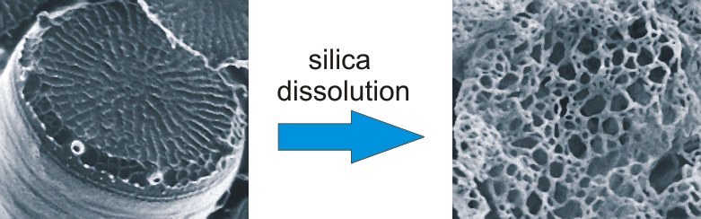
In order to make this discovery, the team dissolved the silica components of diatom shells in an ammonium fluoride solution. The residue appears under the scanning electron microscope to be a delicate, net-like scaffolding. This network resembles the cell wall in form and size and consists of cross-linked fibres with an average diameter of about 25 nanometres. The team carried out a spectroscopic analysis of the fibres, which proved that they are composed mainly of chitin and several previously unknown biomolecules.
“Our results suggest that the chitin-based network structure serves as a supporting scaffold for silica deposition, while the other biomolecules actively influence it,” Brunner explains. “This mechanism is thus analogous to calcium carbonate biomineralization.”
Further work is underway to reveal the function and form of the scaffold materials as well as to investigate other species.
Links: